.svg/1200px-Diagram_of_the_human_heart_(cropped).svg.png)
The sino-atrial node beats at a regular rate of 70 beats/minute.When the electrical impulses reach the atrioventricular node (at the junction between the atria and the ventricles), the impulses will spread rapidly through special fibers from the inter-ventricular septum to the walls of both ventricles, where the muscles are stimulated to contract.

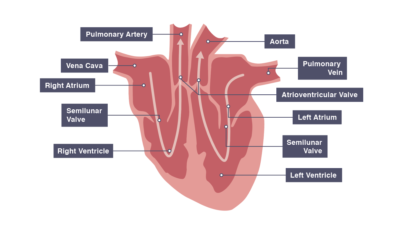
The sino-atrial node sends impulses over the two atria which are then stimulated to contract.The sino-atrial node (the pace maker) is a specialized bundle of thin, cardiac, muscular fibers buried in the right atrial wall, near the connection between the right auricle and the large veins.It has been proven that the heart continues beating regularly even after it has been disconnected from the body and the cardiac nerves. The rhythmic heart beats are actually spontaneous, as they originate from the cardiac tissue itself.The semi-lunar valve can be found where the heart connects with both the aorta and pulmonary artery.The left valve (the bicuspid valve or the mitral valve) has two flaps. The right valve (the tricuspid valve) is made up of three flaps. Blood is permitted to flow only from the atrium into the ventricle, not in the reverse direction. Each atrium is connected to its own ventricle through an opening which is guarded by a valve.The heart is divided longitudinally into two sides by means of muscular walls.They have thick, muscular walls which pump blood through the arteries. The two ventricles: these are the lower two chambers.They have thin walls which receive blood from veins.


The heart is divided into four chambers:.It is enclosed in the pericardium, which protects the heart and facilitates its pumping action. The heart is a hollow muscular organ that lies in the middle of the chest cavity.Keep reading for more detailed A-level Biology revision! Summary The structure of the heart consists of four muscular chambers – the right and left atria and the right and left ventricles – which are separated by valves and blood vessels, allowing for the circulation of blood throughout the body. Which chamber receives deoxygenated blood from the body?.Which chamber of the heart pumps blood to the whole body?.What type of muscles make up the heart?.How many chambers does a human heart have?.


 0 kommentar(er)
